Digital Ultrasound

Digital Ultrasound
The portability and improvement in ultrasound image quality has progressed significantly over the past 10 years. Now being able to follow the healing progress of a bowed tendon or torn ligament has become commonplace. Making measurements and manipulating images to enhance quality as with digital radiography has allowed for quantum leaps in diagnostic abilities. Instead of just doing lower legs other areas are being investigated with excellent results. Diagnostic images of a torn ligament in the back can be seen between vertebrae, a meniscal injury involving a stifle joint can be evaluated and tendon injuries involving the shoulder can be monitored. Recently an exam was done on a horse's eye to diagnose a tumor.
Most lamenesses will require a radiographic evaluation to determine the cause once the problem has been localized. With the advent of digital ultrasound many of these same areas will be evaluated for soft tissue lesions that are not as readily appreciated with x-rays. In fact, it’s rare for a lameness exam not to include both modalities unless it’s quite clear that x-rays or ultrasound by itself are enough to make a conclusive diagnosis.
As with all digital imagery, the software included in the machines as well as available when images are uploaded to a server and sent to other DVMs, consulting radiologists and clients, greatly enhances our ability to focus on and see an area of concern in greater detail.
Another area where ultrasound is frequently used in this practice is to visualize injection into injured structures such as tendons and ligaments, especially with Regenerative Therapies (article on Regenerative Therapies), and also into certain joints and bursae such as a shoulder joint, neck joint facets, Sacro-iliac joints (SI) or a bicipital bursa.
Examples of ultrasound assisted injecting of neck joints:
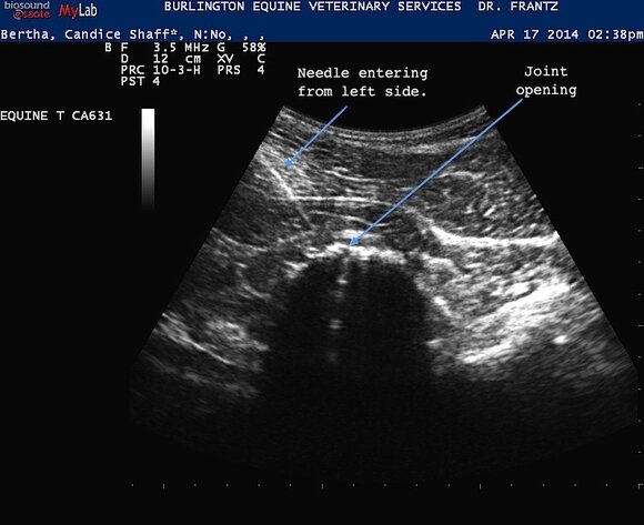

In the above 2 images of a horse’s neck joints, I’m using the ultrasound to direct the needle to the joint openings. to show you what a treatment looks like. This horse had chronic neck arthritis and had multiple treatments over several years by injecting cortisone into the inflamed joints to help manage the limited range of motion and discomfort. Ultrasound allows the visualization of the affected structures and at the same time provides real time images so I can see where the needle will be placed.
Examples of tendon injuries that were diagnosed then monitored for healing:

Image 1 - SDF acute injury
In these 2 images of a Superficial Digital Flexor Tendon injury, the green outlines the tendon and the red indicates the"hole" where the tendon fibers are injured. Over time this area of fiber damage will fill in so that the whole tendon will have a similar pattern as the more white areas.

Image 2 - SDF injury pre-treatment
This is a different horse that horse had a left fore lameness with obvious swelling in the area of his tendon. On the ultrasound exam, there are two things seen above: separation of fibers plus a hole was in the Superficial Digital Flexor Tendon.

Image 3 - SDF injury post-treatment
The horse to the left, received 3 Shockwave treatments over a 6 week period and there was significant improvement in the tendon. The damaged areas had filled in and it was difficult to visualize the previous injury. At this point the horse was able to start controlled walking.
In the above examples seen with the 2nd and 3rd images, this is a typical example on how an ultrasound is used to diagnose then monitor the healing. Typically there will be several ultrasounds done over time especially as the horse progresses from the initial post-treatment phase to doing more active rehab that could include tack walking, ponying then short trot intervals.
Examples of joint related injuries:
The next 2 examples are ultrasounds looking at a stifle specifically at the joints and the menisci. Being able to evaluate a stifle is at least as important, if not more so, than just radiographs. An ultrasound allows us to visualize soft tissue structures such as tendons, ligaments, joint capsules, menisci and cartilage surfaces that are not seen on x-rays unless the damage is more chronic or significant. Article on Stifle Disease

Image 1 Swelling in joint on right side of image (black) plus vertical split in the medial meniscus of a horse with an acute lameness.
In the above image the meniscus, which is the cartilage disk that is on the tibial surface of the stifle joint, can be seen. This is the same type of structure that people have in their knees and can be torn i.e. “a torn meniscus”. Normally this has a triangular shape and a solid grey-white appearance., in this case there’s a vertical split running from the top (external) to the bottom (internal part of the stifle joint). The black space next to this is joint fluid \in one of the stifle joint pouches and is increased indicating joint swelling or effusion. The x-rays for this horse appeared to be normal and he was acutely lame.
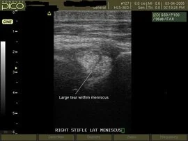
Image 2 An example of a diagonal tear running through the lateral meniscus of a lame horse.
In this image a large tear can be seen running diagonally across the lateral meniscus. This is a significant finding and one that would be consistent with a traumatic injury resulting in lameness. The horse was scheduled to undergo surgical exploration of the joint with arthroscopy.
The ultrasound is used daily with lameness cases and is invaluable for diagnostics, assisting with guided injections and monitoring recovery.