Kissing Spine Disease Article

Kissing Spine Disease - Dorsal Spinous Impingement
Dorsal Spinous Process (DSP) impingement or “Kissing Spines” is a condition recognized as a significant issue for horses. What constitutes the problem is debatable and how to make a conclusive diagnosis can be an elusive process. The following article will discuss the anatomic findings, diagnostic process, therapeutic options and prognosis.
In order to understand the issues behind DSP impingement you have to understand a few anatomical factors. Generally the problem is located in the thoracic section of the vertebral column - the area where the rider sits. Less commonly, involvement of the lumbar vertebrae, the region behind the saddle area, can be the source of the problem. As you can see with the attached picture to the right, the thoracic vertebrae begin with the withers and go through the saddle area.


The part of the vertebrae that we are interested in is the vertical part that projects up and should be evenly spaced from the adjacent process. In the following picture on the left two dorsal spinous processes are seen with the correct relationship along with the attached ribs.
In order to understand the correct relationship between the vertebrae in the back a radiograph is the easiest way to see how the bones sit relative to one another. The following picture is of a combined back radiograph from a horse with normal spacing. The left side of the image starts with the withers and moves down the back to the right showing the lower back. The tall vertical DSP's that make up the withers are narrow and long but are usually not involved with Kissing Spine Disease; instead, it's usually the group of vertebrae behind this area - the thoracic vertebrae, in the area where the rider sits. As you can see the spacing between the DSP‘s is even and there is no significant bone reaction.

The next picture is of an abnormal spine radiograph which clearly depicts kissing spines. As you can see the finger like spinous processes are either touching the adjacent process (or kissing) and in some cases actually overlapping. This horse was having significant issues with under saddle comfort.

Clinical Signs
Horses with this condition can exhibit a range of signs from being asymptomatic, that is exhibiting no abnormal signs, up to having a horse that is unrideable, possibly bucking, refusing to be saddled and/or having behavioral issues even on the ground. The question isn‘t understanding the more obviously affected horse; instead, it‘s the asymptomatic horses. The radiographic changes don’t occur overnight and there are certainly horses that have been in regular work during this time and able to be jumped, be used for dressage and/or western pleasure. This begs the question of how that’s possible as there’s sometimes a disconnect between x-ray findings and clinical signs. Something must change in order for the problem to be evident and something must change again if the problem is going to get under control.
I have heard a variety of complaints from the riders and trainers over the years associated with this condition, the most common one relates to behavior-training issues. Generally the horses may not be overtly lame, but rather exhibiting avoidance behaviors that affect their work such as: refusing to accept bit contact. preferring to travel with their heads up and their backs dropped (not rounded), unwilling to bend one direction or the other, not consistently picking up the correct lead, feeling disconnected or possibly cross cantering. Obviously these signs could be associated with a number of issues for example: stomach ulcers, Lyme disease, tack, training, and/or riders to name a few.
Diagnosis
Making the diagnosis can be a straightforward process in some cases; in others it‘s a process of elimination. The history and clinical signs are particularly beneficial to the Veterinarian. Following this Radiographs are the first line of defense. Digital x-rays allow us to take radiographs of a horse‘s back in a matter of minutes and clearly see if there are issues present as seen in the image below:

Ultrasound is also sometimes employed. Traditionally it‘s been considered a great modality to evaluate soft tissues, but it‘s exceptionally helpful when looking at bone surfaces. While x-rays penetrate bone and show the margins and inside of bones, ultrasound shows a significant amount of detail about the bone surface and soft tissues attached to them. What can‘t be appreciated in some circumstances without ultrasound is back soreness from the ligament that runs over the spine and inserts into these DSP‘s nor the ligaments that are located between the bones. In addition some of the muscles located on the topside of the back called the epaxials can have an important role here. Of these, the Multifidus muscles may be the most important and they can atrophy with chronic muscle issues and/or develop irritations in their tendon attachments to the DSP. All of this can be recognized with ultrasound plus it can show joint issues in the spine that could be another source of back pain.
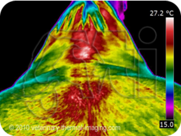
Thermography is also used by me in cases where I want to evaluate heat output which can correlate to inflammation. By using a thermal camera I can visualize areas of the back, legs and even help with saddle fit issues. The more intensely red or white areas correlate with inflammation. The following two pictures demonstrate a more normal back on the left compared to the one on the right with more inflammation in the saddle (thoracic) area as well as over the hips (sacro-iliac) areas.

In cases where there are more questions as to the involvement of the back versus other areas a Nuclear Scan can be done. These are procedures done at referral hospitals. The technique involves administering a medication IV that has the ability to be seen due to its low level of radiation when it collects at an area of increased activity by a camera. The advantage is that, like thermography, they are dynamic exams and show problems in real time as opposed to static exams such as radiographs or ultrasound that show what is currently there, but don‘t necessarily correlate to inflammation or degree or activity. In the picture below the increased whiteness in the thoracic portion of the spine correlates with the Kissing Spine diagnosis.

Treatment
As might be expected treatments can vary. The most important concept is that regardless of the treatment, physical therapy is particularly integral to a successful outcome. What I am referring to here is that the treatments can help manage the soreness short term and even for longer periods, but without instituting a modification in the riding, training and/or tack the problem may continue to affect the horse.
Generally I have found that treatment of the affected areas with injections using a combination of corticosteroids and Sarapin have been beneficial. In most cases that will be my first treatment and often done at the same time the radiographs are taken if the diagnosis is definitive.
Over the years I have treated numerous horses with Shockwave Therapy (SWT) and have also found this effective; in fact, for some horses the benefits are about the same as injections. SWT is also beneficial as a management tool, that is allowing treatment periodically and/or a short time before competitions may decrease the discomfort associated with this chronic condition. Combining injections and SWT may be even more effective for some.
In some of the acute conditions I have effectively used a laser to treat focal areas of inflammation. Laser treatments can treat inflammation of the bone, soft tissues and nerves. Now with our newer Class IV Lasers there’s better penetration and and an alternative to SWT whereby the wave therapies can radiate beyond the sites being treated.
Osphos would be one of the newer medications that I would add to my list. Most of the research with Tildren, a similar drug in the family of medications called Bisphosphonates, had been done in Europe and it has proven benefits for horses with kissing spines. Since then Osphos was used in Europe then it finally went through a testing process and approval in the U.S. with benefits noted in certain bone disease patterns. The essential mechanism is that it decreases the bone destructive process of bone inflammation and “turns on” the bone cells that help to rebuild damaged bone. What is unique with this medication is that its role is not to mask pain, but to improve damaged bone which interrupts the destructive process and improves long term comfort. Based on it’s approval with Kissing Spine Disease and Navicular Disease many of us have effectively extrapolated from there and incorporated it into other bone and joint treatments. As with all medications these drugs are not used without some caveats and these can be discussed with you in detail.
The horse below with kissing spines was unwilling to go forward comfortably, refused jumps and would not collect well was treated with Tildren. Following treatment he was markedly improved and the results lasted longer than when just local injections were used. At this time we would use Osphos an intra-muscular medication vs Tildren which is administered via a catheter IV - much simpler and less costly.

Alternative modalities such as Acupuncture, Chiropractic adjustments, Mesotherapy and/or Massage can also have a role here. I have used them as a first line treatment as well as in a more supportive role. In my opinion if we have a case of significant bone reaction along with kissing spines then the benefits of chiropractic may be minimal and possibly counter productive; however, acupuncture or mesotherapy can reduce the pain and dysfunction. There are a number of individuals in our area that practice equine massage and are frequently recommended by me.
Functional Electrical Stimulation (FES) which I have discussed elsewhere in this website is a different approach than the above, and I find this too, can be particularly helpful when we are trying to manage the muscle spasms and to build back more normal nerve-muscle interactions.

Again Physical Therapy should be considered the essential component for management of this condition. Using exercises that help to build core strength and ones that allow the back to lift are the main concepts. Lunge work often with equipment such as side reins, a Pessoa system, or Equi-Bands are useful tools that promote lifting or flexing of the back and encouraging the horse to engage not just the pelvic (hind end and hip muscles) but also the ones under the spine. Generally I find the Pessoa most useful for this, but the Equi-Bands can work well also.Furthermore there are stretches you can do such as “belly lifts” or “cat scratches” which will help your horse to flex the back and pelvis more and reverse the tendency for the back to be held in an extended or dropped position.
Surgery - new procedure
Interspinous Ligament Desmopathy (ISLD):
This is a surgery that originated in Europe, possibly some initial work was done in France, then later further developed in England. One of the first surgeons to publish information on the procedure and results was Dr. Richard Coomer from Cotts Equine in Wales. Several years ago I contacted Dr. Coomer about some cases that seemed like good candidates for the procedure and asked his opinion. He very graciously replied, reviewed the radiographs and definitely agreed that the new ISLD surgery was appropriate for them. He offered to come over and do some cases with me and while that was appreciated, my goal is to not do surgery, thus I have referred these cases to a surgeon who has done quite a few of these. In the past several years Dr. José Garcia-Lopez from Tufts University Veterinary School has been the surgeon I have referred all of these cases to and we have had excellent results.
The theory behind the surgery is described by Dr. Coomer as follows: "The pain from kissing spines comes from nerve endings present where the ISL sticks on to the bone. Tension and pressure on these nerve endings gives the horse back pain, and causes reflex muscle spasm which pulls the spines even closer and makes it even worse. By cutting the ligament, the nerves stop being stimulated, and the horse experiences a profound improvement in perceived pain. Evidence from France originally suggested that cutting the ligament alone was the important element in successful treatment of kissing spines. We developed this and applied it to the standing horse."
The procedure is done with the horse standing and sedated and a small vertical incision is made adjacent to the involved DSPs on one side then the ISL is cut thus releasing the tension between the two opposing vertebrae. Most of the horses are sent home the next day to rehabilitate at home where they start with hand walking for the first 4 weeks, followed by a period of lunging and turn-out in the 2nd month and if there is appropriate improvement when re-radiographed at 8 weeks they are started back under saddle. So far, unless there have been extenuating circumstances, all of the horses were able to start back with a rider at that point.

In the above picture there are several vertebrae where the spaces between them are mildly narrowed and there is some increased whiteness in some of the edges where the red arrows indicating the ISL attach. Fortunately this horse, a Hunter/Jumper, responded well to medical management. Initially I injected between and adjacent to the affected vertebrae with steroids plus Sarapin. This worked great for the season and he jumped successfully. The following year due to other soundness conditions arising I treated him with Tildren and he had a very positive response with the multiple areas involved. Based on the radiographic signs this horse was a better candidate for medical management vs going to surgery.
For the actual surgery, the surgeon makes a vertical incision that would transect the ISL which is depicted as the area where there are red arrows. The Supraspinous Ligament (SSpL) that runs over the tops of the DSP's is still intact and that stabilizes the vertebrae.
The following two radiographs will demonstrate an actual patient that had the surgery in the summer of 2013. This horse is an 11 year old Irish Sport Horse gelding that had been a jumper for years.


In the above images you can already see some "opening up" of the spaces between the 4 sites. Prior to the surgery this horse became unrideable as he was bucking so much that even low level jumping had become impossible. During the fall once he was started back under saddle all of that had resolved and he was able to start over low jumps and remained very comfortable.
As you may have gathered from this article the simplest part of this discussion is identifying the problem as Kissing Spine Disease from there more diagnostics and treatments may be necessary. While there may be some similarities in presentation, each case is different and is approached that way.
